COVID-19: the difference between the nose and the lung
All claims expressed in this article are solely those of the authors and do not necessarily represent those of their affiliated organizations, or those of the publisher, the editors and the reviewers. Any product that may be evaluated in this article or claim that may be made by its manufacturer is not guaranteed or endorsed by the publisher.
Authors
To the Editor
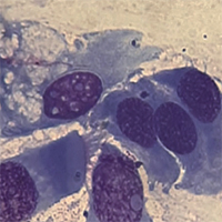
An elegant study reported dysmorphic cells and syncytia in the deceased's lungs for COVID-19. The authors reasonably considered that most of these syncytia-forming cells were pneumocytes, as identified by specific biomarkers. However, cellular dysmorphism and syncytia are pathological features common in other respiratory infections caused by different viruses, including the human respiratory syncytial virus (HRSV) and Epstein-Barr virus (EBV), as correctly documented...


How to Cite

This work is licensed under a Creative Commons Attribution-NonCommercial 4.0 International License.
PAGEPress has chosen to apply the Creative Commons Attribution NonCommercial 4.0 International License (CC BY-NC 4.0) to all manuscripts to be published.
Similar Articles
- H. Halilcolar, S. Yapicioglu, S. Bilaceroglu, Temporal Changes in Lung Cancer: A 10-year Study in a Chest Hospital , Monaldi Archives for Chest Disease: Vol. 69 No. 4 (2008): Pulmonary series
- G.L. Casoni, C. Gurioli, P.N. Chhajed, M. Chilosi, M. Zompatori, D. Olivieri, V. Poletti, The value of transbronchial lung biopsy using jumbo forceps via rigid bronchoscope in diffuse lung disease , Monaldi Archives for Chest Disease: Vol. 69 No. 2 (2008): Pulmonary series
- T. Kontakiotis, N. Manolakoglou, F. Zoglopitis, D. Iakovidis, L. Sacas, A. Papagiannis, A. Mandrali, D. Papakosta, P. Argyropoulou, D. Bouros, Epidemiologic trends in lung cancer over two decades in Northern Greece: an analysis of bronchoscopic data , Monaldi Archives for Chest Disease: Vol. 71 No. 4 (2009): Pulmonary series
- Ali Asghar Hemmati, Soheila Alboghobeish, Akram Ahangarpour, Chronic exposure to high fat diet exacerbates arsenic-induced lung damages in male mice: Possible role for oxidative stress , Monaldi Archives for Chest Disease: Vol. 88 No. 1 (2018)
- Amanda R. Jimenez, Arielle Shaeffer Weiss, Anthony C. Campagna, Use of pulmonary function test demographic data to identify high-risk patients for lung cancer screening , Monaldi Archives for Chest Disease: Vol. 88 No. 1 (2018)
- E.A. Renzoni, Interstitial lung disease in systemic sclerosis , Monaldi Archives for Chest Disease: Vol. 67 No. 4 (2007): Pulmonary series
- S.M. Mirsaeidi, P. Tabarsi, A. Mardanloo, G. Ebrahimi, M. Amiri, P. Farnia, M. Sheikhleslami, V. Bakayev, F. Mohammadi, S.D. Mansouri, M.R. Masjedi, A.A. Velayati, Pulmonary Mycobacterium Simiae infection and HTLV1 infection: an incidental co-infection or a predisposing factor? , Monaldi Archives for Chest Disease: Vol. 65 No. 2 (2006): Pulmonary series
- M. Justine, F. Tahirah, V. Mohan, Health-related quality of life, lung function and dyspnea rating in COPD patients , Monaldi Archives for Chest Disease: Vol. 79 No. 3-4 (2013): Pulmonary series
- Alice Biffi, Giulia Dei, Federica De Giacomi, Anna Stainer, Lorenzo Olmo Parma, Maria Rosa Pozzi, Paola Faverio, Alberto Pesci, Non-specific interstitial pneumonia and features of connective tissue disease: What are the consequences of a different point of view? , Monaldi Archives for Chest Disease: Vol. 88 No. 3 (2018)
- I.P. Neuringer, S.H. Randell, Lung Stem Cell Update: Promise and Controversy , Monaldi Archives for Chest Disease: Vol. 65 No. 1 (2006): Pulmonary series
You may also start an advanced similarity search for this article.
