See how this article has been cited at scite.ai
scite shows how a scientific paper has been cited by providing the context of the citation, a classification describing whether it supports, mentions, or contrasts the cited claim, and a label indicating in which section the citation was made.
Convex probe endobronchial ultrasound guided transbronchial/transoesophageal fine needle aspiration (C-EBUS-TBNA/EUS-B FNA) of pleural lesions: A single center experience and review of literature
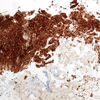
The evaluation of pleural diseases has been well established. If pleurocentensis is non-diagnostic, the second investigation depending upon availability could be either closed pleural biopsy or image guided pleural biopsy or thoracoscopic pleural biopsy (medical or surgical). Pleural disease presenting as thickness/mass/nodule in the mediastinum is difficult to access through ultrasound or computed tomography and will need thoracoscopy. Thoracoscopy is an invasive procedure which can be difficult to perform in localized mediastinal pleural disease without effusion or poor health condition not suitable for general anesthesia. An alternative method that can be utilized is sampling of pleural lesion through convex probe endobronchial ultrasound (CEBUS) either through the central large airways or from esophagus if the lesions are in proximity. We present our center’s experience in diagnosing pleural lesion using C-EBUS in 4 patients along with review of the literature.
Downloads
Citations


How to Cite
PAGEPress has chosen to apply the Creative Commons Attribution NonCommercial 4.0 International License (CC BY-NC 4.0) to all manuscripts to be published.