0
0
0
0
Smart Citations
0
0
0
0
Citing PublicationsSupportingMentioningContrasting
View Citations

See how this article has been cited at scite.ai

scite shows how a scientific paper has been cited by providing the context of the citation, a classification describing whether it supports, mentions, or contrasts the cited claim, and a label indicating in which section the citation was made.

A case of B.1.1.7 SARS-CoV-2 UK strain with an atypical radiological presentation

Authors

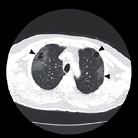
The new UK strain was first described in December 2020. It was seen for the first time in Turkey in February 2021. It is not yet known whether the new strain has different CT patterns compared to the classical type. We present a 68-years-old male patient with an atypical CT presentation in which GGOs are gathered around the areas of paraseptal emphysema accompanied by CT and clinical findings. This involvement is an unexpected pattern because of the atypical distribution of the GGO.

Tenda ED, Yulianti M, Asaf MM, et al. The importance of chest CT scan in COVID-19. Acta Med Indones 2020;52:68-73.
Ufuk F, Savaş R. Chest CT features of the novel coronavirus disease (COVID-19). Turk J Med Sci 2020;50:664–78.
Kirby T. New variant of SARS-CoV-2 in UK causes surge of COVID-19. Lancet Respir Med 2021;9:20-1. DOI: https://doi.org/10.1016/S2213-2600(21)00005-9
Salehi S, Abedi A, Balakrishnan S, Gholamrezanezhad A. Coronavirus disease 2019 (COVID-19): A systematic review of imaging findings in 919 patients. AJR Am J Roentgenol 2020;215:1–7. DOI: https://doi.org/10.2214/AJR.20.23034
Kupferschmidt K. Fast-spreading U.K. virus variant raises alarms. Science 2021;371:9-10. DOI: https://doi.org/10.1126/science.371.6524.9
Takahashi M, Fukuoka J, Nitta N, et al. Imaging of pulmonary emphysema: a pictorial review. Int J Chron Obstruct Pulmon Dis 2008;3:193-204. DOI: https://doi.org/10.2147/COPD.S2639
Garg M, Prabhakar N, Gulati A, et al. Spectrum of imaging findings in pulmonary infections. Part 1: Bacterial and viral. Pol J Radiol 2019;84:205-13. DOI: https://doi.org/10.5114/pjr.2019.85812
Franquet T, Chung JH. Imaging of pulmonary infection. In: Hodler J, Kubik-Huch RA, von Schulthess GK, editors. Diseases of the chest, breast, heart and vessels 2019-2022: Diagnostic and interventional imaging [Internet]. Cham: Springer; 2019. Chapter 7. DOI: https://doi.org/10.1007/978-3-030-11149-6_7
Morinet F, Parent M, Bergeron C, et al. Oxygen and viruses: a breathing story. J Gen Virol 2015;96:1979-82. DOI: https://doi.org/10.1099/vir.0.000172
Koo HJ, Lim S, Choe J, et al. Radiographic and CT features of viral pneumonia. Radiographics 2018;38:719-39. DOI: https://doi.org/10.1148/rg.2018170048
Hamad AMM, El-Saka HA. Post COVID-19 large pneumatocele: clinical and pathological perspectives. Interact CardioVasc Thor Surg 2021;ivab072. DOI: https://doi.org/10.1093/icvts/ivab072
Zhou S, Wang Y, Zhu T, Xia L. CT Features of coronavirus disease 2019 (COVID-19) pneumonia in 62 patients in Wuhan, China. AJR Am J Roentgenol 2020;214:1287-94. DOI: https://doi.org/10.2214/AJR.20.22975
Ufuk F, Savaş R. Chest CT features of the novel coronavirus disease (COVID-19). Turk J Med Sci 2020;50:664-78. DOI: https://doi.org/10.3906/sag-2004-331
Han X, Fan Y, Alwalid O, et al. Six-month follow-up chest CT findings after severe COVID-19 pneumonia. Radiology 2021;299:E177-6. DOI: https://doi.org/10.1148/radiol.2021203153

How to Cite

Doğan, Emrah, Utku Tapan, Özge Oral Tapan, Fatih Alaşan, Sabri Serhan Olcay, and Tuğba Çınar Olcay. 2021. “A Case of B.1.1.7 SARS-CoV-2 UK Strain With an Atypical Radiological Presentation”. Monaldi Archives for Chest Disease 91 (4). https://doi.org/10.4081/monaldi.2021.1840.

Similar Articles

<< < 2 3 4 5 6 7 8 9 10 11 > >> 

You may also start an advanced similarity search for this article.