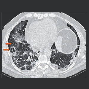
Bull’s eye sign – A diagnostic clinch in COVID-19 pneumonia

Published: September 28, 2021
Abstract Views: 1265
PDF: 392
Publisher's note
All claims expressed in this article are solely those of the authors and do not necessarily represent those of their affiliated organizations, or those of the publisher, the editors and the reviewers. Any product that may be evaluated in this article or claim that may be made by its manufacturer is not guaranteed or endorsed by the publisher.
All claims expressed in this article are solely those of the authors and do not necessarily represent those of their affiliated organizations, or those of the publisher, the editors and the reviewers. Any product that may be evaluated in this article or claim that may be made by its manufacturer is not guaranteed or endorsed by the publisher.
Similar Articles
- F. Blasi, S. Aliberti, Pneumonia: how important are local epidemiology and smoking habits? , Monaldi Archives for Chest Disease: Vol. 65 No. 1 (2006): Pulmonary series
- G. Caramori, D. Artioli, G. Ferrara, R. Cazzuffi, C. Pasquini, M. Libanore, V. Guardigni, I. Guzzinati, M. Contoli, R. Rossi, R. Rinaldi, C. Contini, A. Papi, Severe pneumonia after intravesical BCG instillation in a patient with invasive bladder cancer: case report and literature review , Monaldi Archives for Chest Disease: Vol. 79 No. 1 (2013): Pulmonary series
- Vittorio Palmieri, Antonio Palermo, Patients’ self-evaluation of symptoms, signs and compliance to therapy for heart failure surveillance: A pilot study on identification of worsening heart failure , Monaldi Archives for Chest Disease: Vol. 88 No. 2 (2018)
- M. Messina, N. Scichilone, F. Guddo, V. Bellia, Rapidly progressive organising pneumonia associated with cytomegalovirus infection in a patient with psoriasis , Monaldi Archives for Chest Disease: Vol. 67 No. 3 (2007): Pulmonary series
- William Newmarch, Angelica Puopolo, Madina Weiler, Brian Casserly, Hamman-Rich syndrome: a forgotten entity , Monaldi Archives for Chest Disease: Vol. 87 No. 1 (2017)
- S.W. Yan, C.F. Wong, P.C. Wong, C.F. Cheung, Bronchiolitis Obliterans Organising Pneumonia (BOOP) in a lung cancer patient after lobectomy , Monaldi Archives for Chest Disease: Vol. 63 No. 1 (2005): Pulmonary series
- R.M. Jones, A. Dawson, E.N. Evans, N.K. Harrison, Co-existence of organising pneumonia in a patient with Mycobacterium Avium Intracellulare pulmonary infection , Monaldi Archives for Chest Disease: Vol. 71 No. 2 (2009): Pulmonary series
- R.A. Stolarek, M. Kasielski, J. Rysz, P. Bialasiewicz, D. Nowak, Differential effect of cigarette smoking on hydrogen peroxide and thiobarbituric acid reactive substances exhaled in patients with community acquired pneumonia , Monaldi Archives for Chest Disease: Vol. 65 No. 1 (2006): Pulmonary series
- Ravi Manglani, Moshe Fenster, Theresa Henson, Ananth Jain, Neil Schluger, Clinical characteristics, imaging, and lung function among patients with persistent dyspnea of COVID-19: a retrospective observational cohort study , Monaldi Archives for Chest Disease: Early Access
- Elpida Skouvaklidou, Ioannis Neofytou, Maria Kipourou, Konstantinos Katsoulis, Persistent unilateral diaphragmatic paralysis in the course of Coronavirus Disease 2019 pneumonia: a case report , Monaldi Archives for Chest Disease: Vol. 93 No. 4 (2023)
<< < 1 2 3 4 5 6 7 8 9 10 > >>
You may also start an advanced similarity search for this article.

 https://doi.org/10.4081/monaldi.2021.1908


