Early diagnosis of cardiomyopathies by cardiac magnetic resonance. Overview of the main criteria

All claims expressed in this article are solely those of the authors and do not necessarily represent those of their affiliated organizations, or those of the publisher, the editors and the reviewers. Any product that may be evaluated in this article or claim that may be made by its manufacturer is not guaranteed or endorsed by the publisher.
Authors
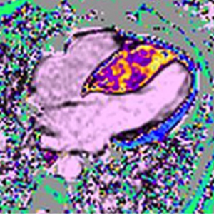
Cardiomyopathies (CMPs) are diseases of the heart muscle. They include a variety of myocardial disorders that manifest with various structural and functional phenotypes and are frequently genetic. Myocardial disease caused by known cardiovascular causes (such as hypertension, ischemic heart disease, or valvular disease) should be distinguished from CMPs for classification and management purposes. Identification of various CMP phenotypes relies primarily upon echocardiographic evaluation. In selected cases, cardiac magnetic resonance imaging (CMR) or computed tomography may be useful to identify and localize fatty infiltration, inflammation, scar/fibrosis, focal hypertrophy, and better visualize the left ventricular apex and right ventricle. CMR imaging has emerged as a comprehensive tool for the diagnosis and follow-up of patients with CMPs. The accuracy and reproducibility in evaluating cardiac structures, the unique ability of non-invasive tissue characterization and the lack of ionizing radiation, make CMR very attractive as a potential “all-in-one technique”. Indeed, it provides valuable data to confirm or establish the diagnosis, screen subclinical cases, identify aetiology, establish the prognosis. Additionally, it provides information for setting a risk stratification (based on evaluation of proved independent prognostic factors as ejection fraction, end-systolic-volume, myocardial fibrosis) and follow-up. Last, it helps to monitor the response to the therapy. In this review, the pivotal role of CMR in the comprehensive evaluation of patients with CMP is discussed, highlighting the key features guiding differential diagnosis and the assessment of prognosis.
How to Cite

This work is licensed under a Creative Commons Attribution-NonCommercial 4.0 International License.


